KOR
KOR ENG
ENG


Home / 제품소개 / 상세페이지
")

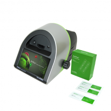
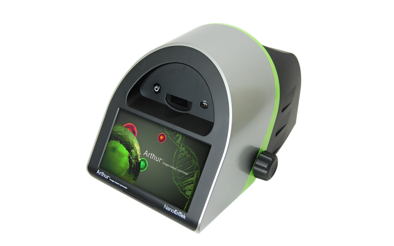
Fluorescence Cell Counter
- Accurate cell counting & analysis
- Various & fast analysis assay tools
- Rapid set up & user friendly interface
- No need for maintenance


It takes Arthur™ 10 seconds to 2 minutes to complete the assay with only 20 μL of sample.
-Advanced accurate cell counting & analysis
- Various analysis assay tools (GFP/RFP expression, apoptosis, cell viability, cell cycle and cell counting)
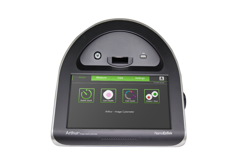
Assays and applications
Arthur™ features two fluorescent channels (green & red) as well as bright field.
Arthur™ performs a broad range of assays : cell counting & viability, GFP&RFP expression, Annexin V apoptosis analysis, and cell cycle analysis.
Cell viability assay
Figure1. Comparison of cell viability in two platforms
Head shocked cells(proteins) were stained with Propidium Iodide (PI) and percent of viable cells of in the total population
were analyzed by Arthur™(A) and competitor's product F (B) with two different cell types, Jurkat and U-2 OS.
In all cases, the fluorescence threshold setting was confirmed visually in the image of the Arthur™.
For each cell line, viability results from the Arthur™ were consistent with the results obtained from the competitor's product F.


GFP expression assay
Figure2. Comparison of GFP expression profile in two platforms
U-2 OS cells were transfected using the Neon transfection system (from Life Technologies) and 0.25 μg of the five different GFP constructs
and then analyzed on Arthur™ and competitor's product F to determine transfection efficiency.
The histograms in panels A (Arthur™) and C (competitor's product F) show the green fluorescence intensities and counts for the transfected populations.
The percentages of GFP-expressing cells detected with Arthur™ were consistent with the results from the competitor's product F.
'Colored circles' option makes it easy to identify gated cells (Panel B); colored circles represent
: cell expressing GFP (green circles), non-GFP-expressing cells (blue circles).
The image in panel D shows the GFP expressing cells before trypsinization observed with a conventional fluorescence microscope.


Annexin V apoptosis
Figure3. Arthur™ allows confirmation of apoptosis assay using the visual display of the samples.
Following 18 hour-treatment of Staurosporine, HeLa cells were stained with apoptosis kit and subjected to Arthur™ and competitor's product F.
Apoptosis Kit contains an annexin V-Alexa Fluor 488 conjugate and PI to differentiate
Live (Annexin V-negative / PI-negative), Dead (PI-positive) and Apoptotic (Annexin V-positive / PI-negative) cells.


Cell cycle assay
Figure4. Comparison of cell cycle profiles in two platforms : Arthur™ and competitor's Product F
HeLa cells were treated with Nocodazole (Microtubule-Destabilizer) or Epothilone B (Microtubule-Stabilizer) to arrest cells in G2/M phase.
Cells were fixed with 70% ethanol and stained with Propidium Iodide (PI).
The histograms show the percentages of G0/G1 phase, S phase and G2/M phase of cell cycle analysis for
drug treated HeLa cells on Arthur™ (A) and competitor's product F (B).
The cell cycle analysis of Arthur™ was highly consistent with the results obtained from competitor's product F


Visit NanoEntek blog now!
Want to learn more about cell counting and cell therapy?
Visit to find out more!

-
Product name / Cat. No.
Arthur™ / AT1000
-
Cell measurement range
1 × 10E5–1 × 10E7 cells/mL
-
Cell size range
5–60 μm
-
Sample volume
20 μL
-
Excitation
Green channel LED : 458 ± 20 nm & Red channel LED : 530 ± 20 nm
-
Filters
Green channel : 466/40 EX, 495 LP Di, 525/50 EM & Red channel : 543/22 EX, 580 LP Di, 585 LP EM
-
Camera
1.3 Mega pixels, 4 X objective , 4 X or 16 X digital zoom
-
Operating power
100–240 V, 2.5 A, 120 W
-
Dimension
290 mm (W) x 440 mm (D) x 290 mm (H)
-
Weight
8.7 kg
-
Operating environment
5–40 ℃
-
Optics
3 Channels (bright field, green fluorescence, red fluorescence)
-
Counting time
10 sec–2 min
Related downloads Total 2
-
Arthur_brochure_NanoEntek

-
Arthur_user_manual_NanoEntek


ExTransfection™ Protocol Library
ExTransfection™ provides open protocols that make it easy for users to set up and run experiments. Its 24-well optimization method helps users to optimize transfection protocols easily and quickly.
ExTransfection™ also offers a pre-programmed optimization protocol that helps an easy and quick optimization of electrical parameters for both adherent and suspended cells.