KOR
KOR ENG
ENG


Home / 제품소개 / 상세페이지


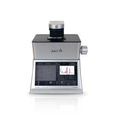
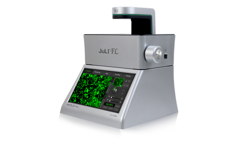
Live fluorescent cell movie analyzer
- Incubator-compatible
- Time-lapse image capturing and movie making
- GFP or RFP expression level checking
- Compact size optimized for a cell culture incubator
- Dual system for data comparison (optional)

Meet JuLI™ FL to meet your needs
for live cell imaging
The compact design allows you to install the system in your cell-culture incubator easily.

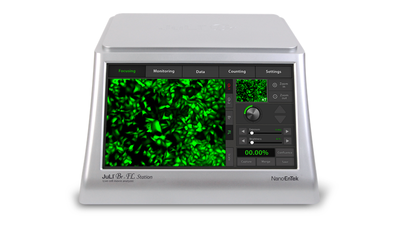
Time-lapse image capture & movie making
Cell-growth images were captured for 63 hours with 20 minutes interval in U-2 OS GFP & RFP stable cell line.

Real time cell growth curve
HeLa cells growth were observed 40 hours with 10 minutes intervals
and JuLI™ FL analyzed monolayer confluence.
For apoptosis assays, experimental group (channel 2) was treated by Staurosporine.

Cell migration (wound healing) assay
U-2 OS stable clees were incubated for 100 hours after scratch.
JuLI™ FL calculated the confluence with growth of surface unfarmed automatically.

Applications
Cell growth monitoring / Cell culture quality control / Cell migration assay
Proliferation assay / Cell confluence detection / Cell based assay optimization / Cell viability & counting

Visit NanoEntek blog now!
Want to learn more about cell counting and cell therapy?
Visit to find out more!

-
Power
AC 100–240V, 50/60 Hz
-
Weight
Scope : 4.5kg, Station 3.2Kg
-
Objective lens
4 X & digital zoom
-
Image resolution
1280 X 960 pixels (1.3M)
-
Display
10.1
-
Data storage
Hard drive (320 GB), USB drive (4 GB)
-
Operation temperature
5–40 ℃
-
Maximum relative humidity
20–95 %
-
Counting time
< 10 sec/test
-
Cell measurement range
1X10E4–1X10E7 cell/μL
-
Optimal measurement range
1X10E5–4X10E6 cell/μL
-
Cell size range
5–60 μm
-
Sample volume
10 μL
-
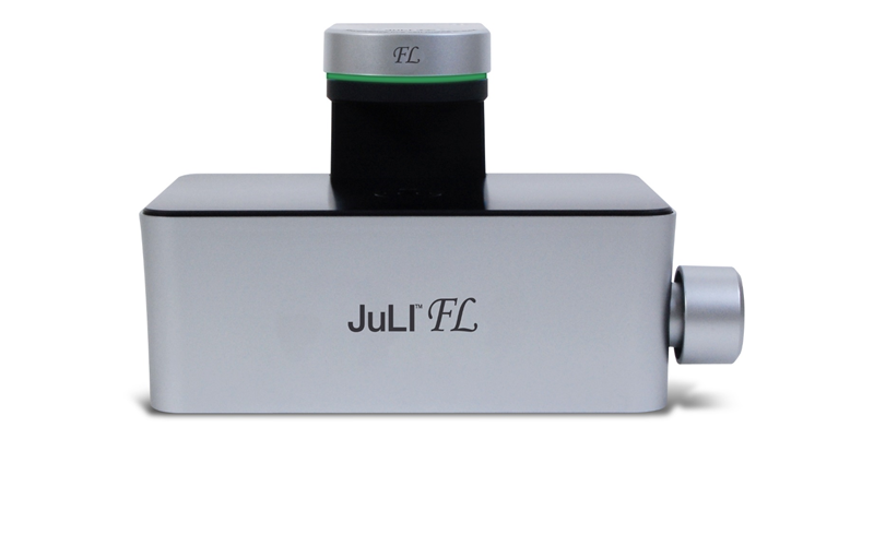
Size
Scope 300 (W) X 190(L) X 188 (H) mm & Station 282 (W) X 285 (L) X 160 (H) mm
-
Data format
JPEG, TIFF, BMP, PNG (image), AVI (movie), CSV (raw data)
-
Light source
GFP channel (JULI–FLG04): Blue LED & RFP channel (JULI–FLG04): Green LED
-
Optical filter
GFP channel (JULI–FLG04): Excitation 466/40nm, Emission: 525/50nm & RFP channel (JULI–FLG04): Excitation 525/50 nm, Emission 580LP
Related downloads Total 2
-
JuLI_FL_Brochure_NanoEntek

-
JuLI_FL_User_manual_NanoEntek
-
LS PRODUCT / JuLI™ FL
It dims out or gets brighter over time when recording (the scope is placed inside an incubator).

 If the scope is not enough warmed up, images may look gray due to the temperature difference between the scope and inside the incubator. Please place the scope in the incubator for an hour for warm-up before use.
 -
LS PRODUCT / JuLI™ FL
Data is not saved into a USB drive when other file formats than JPEG chosen.
1. Please make sure the software is most up to date. Please contact us for more information on software updates.2. The latest software information : JuLI FL S/W (Ver.1.0.5.6), JuLI Br S/W (Ver.1.3.7.5) -
LS PRODUCT / JuLI™ FL
I see debris in the images taken.
1. Please check if there is any debris on the objective lens and softly wipe the objective using cotton swabs. If the problem persists, please contact us.2. The incubator environment may leave the scope vulnerable to erosion or mold if placed inside for an extended amount of time. Please store the scope outside an incubator when not in use.


ExTransfection™ Protocol Library
ExTransfection™ provides open protocols that make it easy for users to set up and run experiments. Its 24-well optimization method helps users to optimize transfection protocols easily and quickly.
ExTransfection™ also offers a pre-programmed optimization protocol that helps an easy and quick optimization of electrical parameters for both adherent and suspended cells.